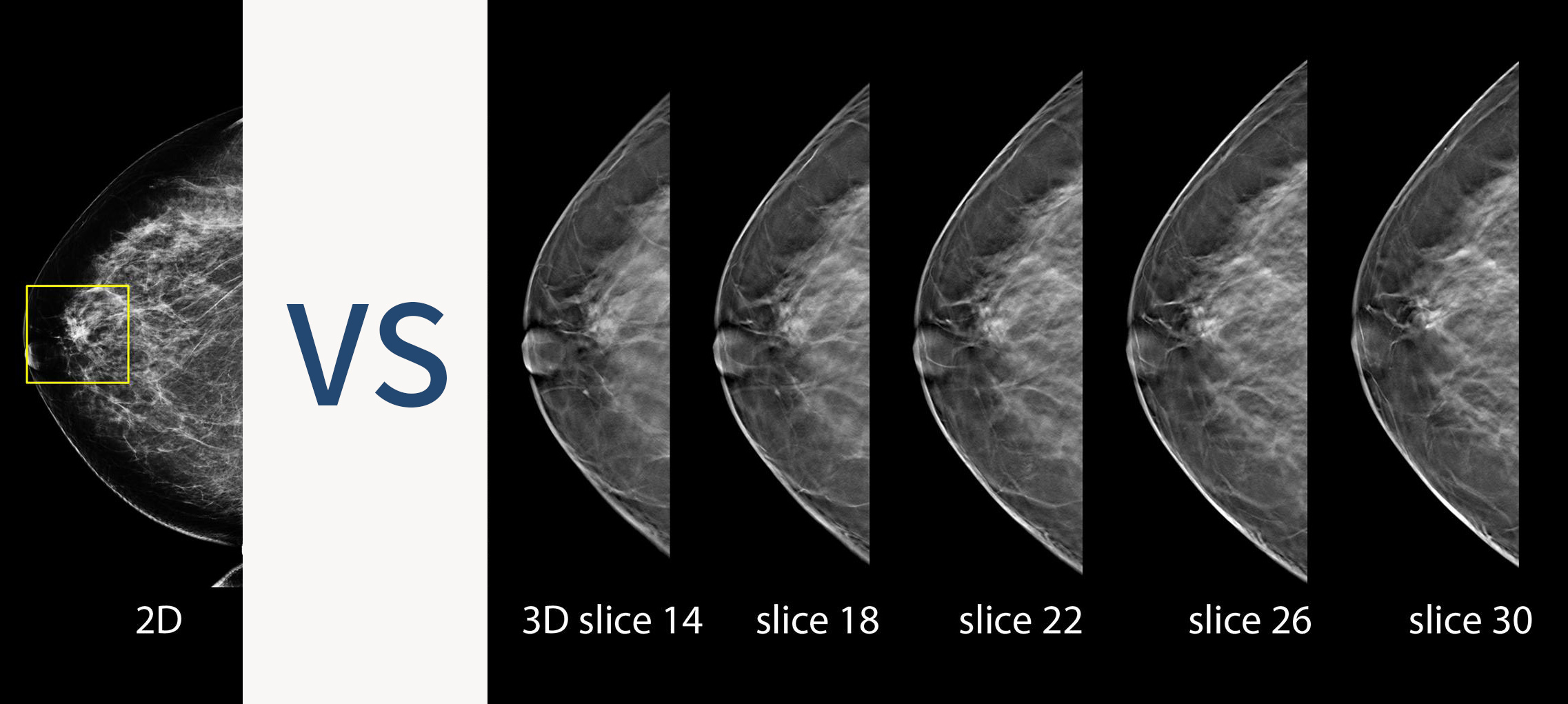
Breast Tomosynthesis uses low-dose X ray system and advanced computing technology to create multiple cross-sectional images of breast tissue. It is also known as 3D Mammography because it makes use of a series of 2D images to reconstruct 3D image of your breast. It is effective for premenopausal women with dense breast tissue. Comparing with conventional mammography, this method increases the chances of finding small and invasive breast cancers tumors by 41%*.
✔ It reduces overlapping of breast tissue, thus enhancing visibility of abnormalities.
✔ The radiation dose is slightly higher than 2D Mammography but it is considered as a reasonable trade-off for improved cancer detection.
( ![]() Click the image to enlarge ) Image comparison between 2D &3D Mammography
Click the image to enlarge ) Image comparison between 2D &3D Mammography
Image source: Hologic, Inc.
Decision between 2D and 3D Mammography depends on your medical history, breast density and specific clinical recommendations. You may consult your doctors for further information.
Reference :
* Friedewald, S. M., Rafferty, E. A., Rose, S. L., Durand, M. A., Plecha, D. M., Greenberg, J. S., Hayes, M. K., Copit, D. S., Carlson, K. L., Cink, T. M., Barke, L. D., Greer, L. N., Miller, D. P., & Conant, E. F. (2014). Breast cancer screening using tomosynthesis in combination with digital mammography. JAMA, 311(24), 2499–2507.


