



What Is an Ultrasound Scan?
Ultrasound Imaging Principles: Non-Invasive and Non-Radiation Imaging
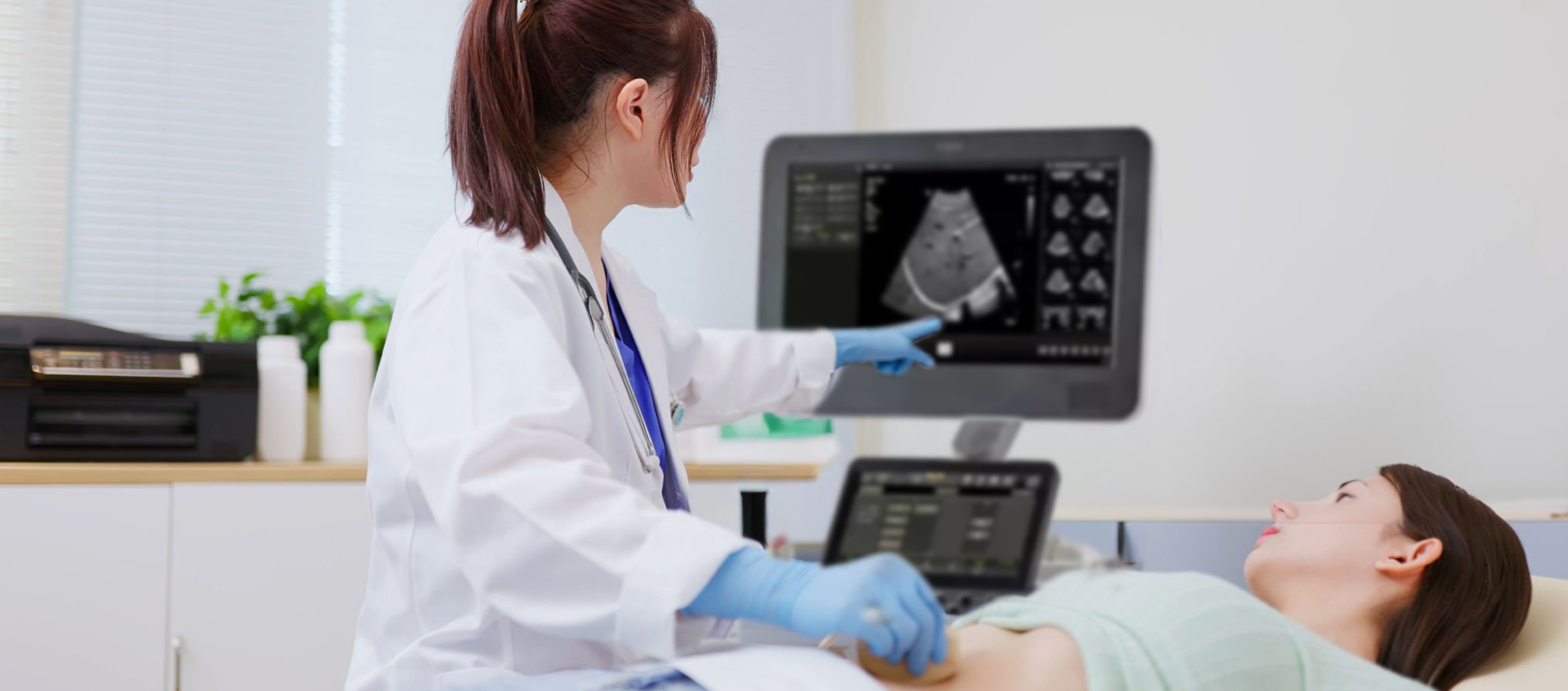
An ultrasound scan is a medical imaging technique that uses high-frequency sound waves (greater than 20,000 Hz, beyond the range of human hearing) to examine internal structures of the body. These sound waves can pass through the skin and tissues. By using high-resolution imaging technology, together with computer-based high-speed calculations and signal processing, ultrasound imaging can instantly create images showing the shape, size, and texture of internal organs.
Because ultrasound uses no radiation, doctors can also use it to monitor a fetus's growth and development inside the mother.
Advantages and Limitations of Ultrasound Scanning
During an ultrasound scan, doctors can observe organ movement in real time—for example, the beating of the heart or blood flow in blood vessels. It is not only considered safe but is also generally comfortable and painless.
However, ultrasound is not all-powerful. Sound waves have difficulty passing through bone or air. Therefore, organs surrounded by bone (such as the brain) or organs filled with air (such as the lungs and intestines) may be harder to image clearly. For certain scan types, patients must follow specific preparation instructions—such as fasting or having a full bladder—to reduce interference from air or gas.
Common Ultrasound Scan Types
Upper Abdominal Ultrasound (Liver, Gallbladder, Spleen, Pancreas, Kidneys)
Pelvic and Lower Abdominal Ultrasound (Uterus, Ovaries, Prostate)
Breast Ultrasound (Breast Lumps, Gland Evaluation)
Neck and Thyroid Ultrasound
Vascular Ultrasound (e.g., Carotid Arteries, Deep Veins)
Pre-Scan Preparation: Special Requirements for Different Body Areas
To ensure clear images, patients must complete the appropriate preparations before the scan. In general, the scan takes about 20 to 30 minutes. On the day of the examination, wear simple, loose clothing. You may need to uncover the relevant area or change into an examination gown.
Whole Abdomen Ultrasound: Key Preparation Tips
If you are having both upper abdominal and pelvic scans, you must follow both preparation requirements: fasting and having a full bladder.
Upper Abdomen Ultrasound: Why You Need to Fast for 6–8 Hours
Upper abdominal scanning typically requires strict fasting (usually 6–8 hours, and you may drink a small amount of water).
- To prevent gallbladder contraction
Eating fatty or protein-rich foods can stimulate the release of cholecystokinin (CCK) from the duodenum. This causes the gallbladder to contract and release bile. After contraction, the gallbladder wall can become thicker and more wrinkled, making it harder for the doctor to accurately assess the gallbladder wall thickness or detect tiny gallstones and polyps. Fasting allows the gallbladder to fill with bile, stretching the wall and creating a clearer liquid interface for observation. - To reduce interference from gastrointestinal gas
During eating, swallowed air and gases produced during digestion can accumulate in the stomach and intestines. Because ultrasound cannot penetrate gas well, these gases form a “sound barrier,” blocking views of structures behind the stomach—such as the pancreas and the abdominal aorta—and can significantly reduce diagnostic accuracy.
Pelvic/Prostate Ultrasound: Why You Must Drink Water and Hold Urine
Before undergoing an abdominal pelvic ultrasound, patients are usually asked to drink 4 to 5 cups of water to fill the bladder.
- To move the intestines out of the way
A full bladder acts like an “inflated balloon,” pushing gas-filled intestines upward and reducing obstacles that may otherwise block the view of the uterus, ovaries, or prostate. - To create a better acoustic window
Urine is a good medium for conducting ultrasound waves. Ultrasound can pass easily through a bladder filled with urine and reach the pelvic organs behind it, producing clearer images of deeper structures.
Other Areas (Breast, Thyroid, Neck, etc.): No Special Preparation is Required
Besides the abdominal and pelvic scans above, if you are getting an ultrasound of the breast, thyroid, neck, or other areas, you usually do not need special pre-scan preparation. Just arrive on time and wear comfortable clothing.
Ultrasound Scan Process
Pre-Scan Preparation and Changing Clothes
To make it easier to scan the abdomen or neck, patients may need to change into a loose examination gown and remove any jewelry.
During the Scan: The Role of Conductive Gel and What It Feels Like
During the exam, the doctor will apply a layer of water-based gel to the skin. Because air blocks high-frequency sound waves, the gel removes the air layer between the probe and the skin. This helps the ultrasound waves transmit into the body properly, enabling clear imaging.
The gel is usually cool to the touch but is not irritating. After the scan, you can wipe it off with tissue.
After the Scan: When Can You Get the Report?
The scan typically takes about 20 to 30 minutes.
After the exam, patients can usually return right away to normal eating and daily activities (unless the scan includes an interventional needle procedure). Written reports are typically available within a few days.
Ultrasound Safety Information and Common Questions
Are There Any Side Effects? Is It Suitable for Pregnant Women and Children?
Ultrasound imaging does not use X-rays or any radioactive materials. After the scan, patients can immediately resume normal activities. It does not cause cell mutations or increase cancer risk. It is suitable for pregnant women for prenatal checks and for children's organ evaluations.
How Does Ultrasound Differ from CT/MRI?
- Ultrasound: No radiation, real-time imaging, and generally lower cost. Limited penetration as it cannot pass well through bone or air.
- CT (Computed Tomography): Fast imaging and clear images for lung and bone conditions, but involves radiation exposure.
- MRI (Magnetic Resonance Imaging): Provides excellent imaging for soft tissues and no radiation, but scans take longer and may not be suitable for people with metal implants.
Can Ultrasound Replace a Biopsy?
Ultrasound can effectively detect lumps and distinguish some physical characteristics (such as whether a lesion is cystic or solid). However, imaging alone cannot definitively confirm whether a lump is malignant. If ultrasound finds a suspicious lesion, doctors may recommend an ultrasound-guided needle biopsy to collect tissue cells for laboratory testing, which provides a more accurate diagnosis.
If you want, I can also format this into a polished, publication-ready article layout (with consistent medical tone and shorter paragraphs).
Can Ultrasound Apply in Therapeutic purpose
Ultrasound technology for cancer treatment is mainly divided into High-Intensity Focused Ultrasound (HIFU) and the innovative Histotripsy. These methods are non-invasive, incisionless treatments that precisely target and destroy cancer cells while minimizing damage to the surrounding healthy tissue. They are suitable for specific patients with liver, kidney, and prostate tumors.
Learn about other professional services:


